
Surgical Margin Evaluation of Clear Cell Renal Cell Carcinoma (ccRCC) Using Ddesorption Electrospray Ionization Mass Spectrometry Imaging (DESI-MSI)
Vijaya Lakshmi Kanchustambham
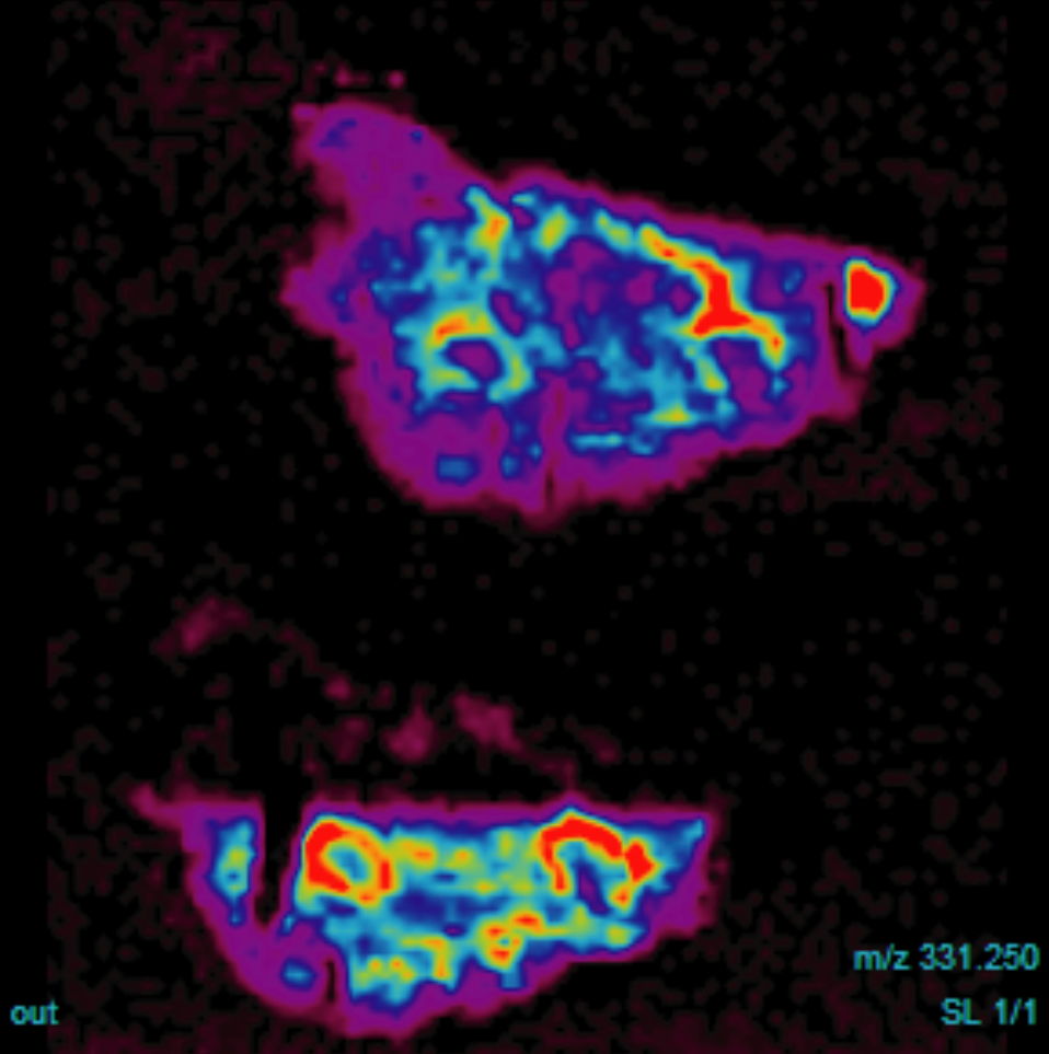
Figure 1. Chemical map (m/z = 331.250) of metabolic signatures in ccRCC positive surgical margins probed by DESI-MSI
One of the great challenges in tumor-progression-free survival after surgical resection of solid tumors is to distinguish the delicate boundary between positive and negative surgical margins. Conventional histopathologic evaluation of surgical margins using intraoperative frozen sections (IFS) is time-consuming, expensive, and susceptible to artefacts. The demand for fast and accurate method for cancer detection and diagnosis led to the development of both ex-vivo and in-vivo molecular imaging techniques. DESI-MSI is an ambient ionization mass spectrometry imaging technique which offers unique potential for the chemical map of clinical biomarkers.1 Using DESI-MSI, we image both normal and cancer kidney tissue sections and identify the metabolites and lipid signatures that are unique to the cancer abnormal metabolism. The classification and validation of potential biomarkers is performed by the statistical method least absolute shrinkage and selector operator (LASSO) of the mass spectrometry imaging data from paired surgical margins of patients.